

CLICK PHOTO TO ENLARGE
Case #:
6
Presented by:
Dr Benjamin Yongcheng Tan
Department of Pathology, Singapore General Hospital
Clinical History:
A 61-year-old Asian female with hemodialysis-dependent end-stage renal failure and a significant medical history of hypertension, diabetes mellitus, deep vein thrombosis, and pulmonary embolism presented with progressive enlargement of her left arm and breast. Magnetic resonance imaging revealed left axillary vein thrombosis extending to the left subclavian vein, without significant axillary lymphadenopathy. She had a previous episode of left upper limb cellulitis, which resolved after inpatient management. Owing to failure of conservative treatment, which included a period of compression garment therapy, the patient elected for a mastectomy.
Pathology:
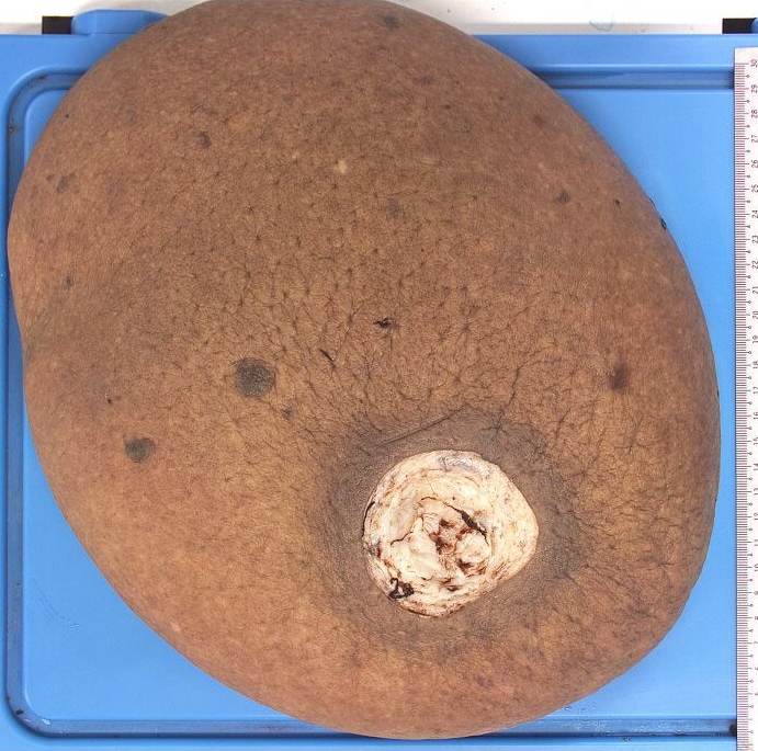
Macroscopic findings: The left simple mastectomy specimen measured 31cm x 30cm x 9cm and weighed 4.1kg. The overlying skin showed generalised brawny discolouration. Sectioning the breast revealed an edematous appearance with copious clear fluid oozing from the cut surfaces. A well-circumscribed nodule measuring 1.1cm in diameter was identified within the breast substance.
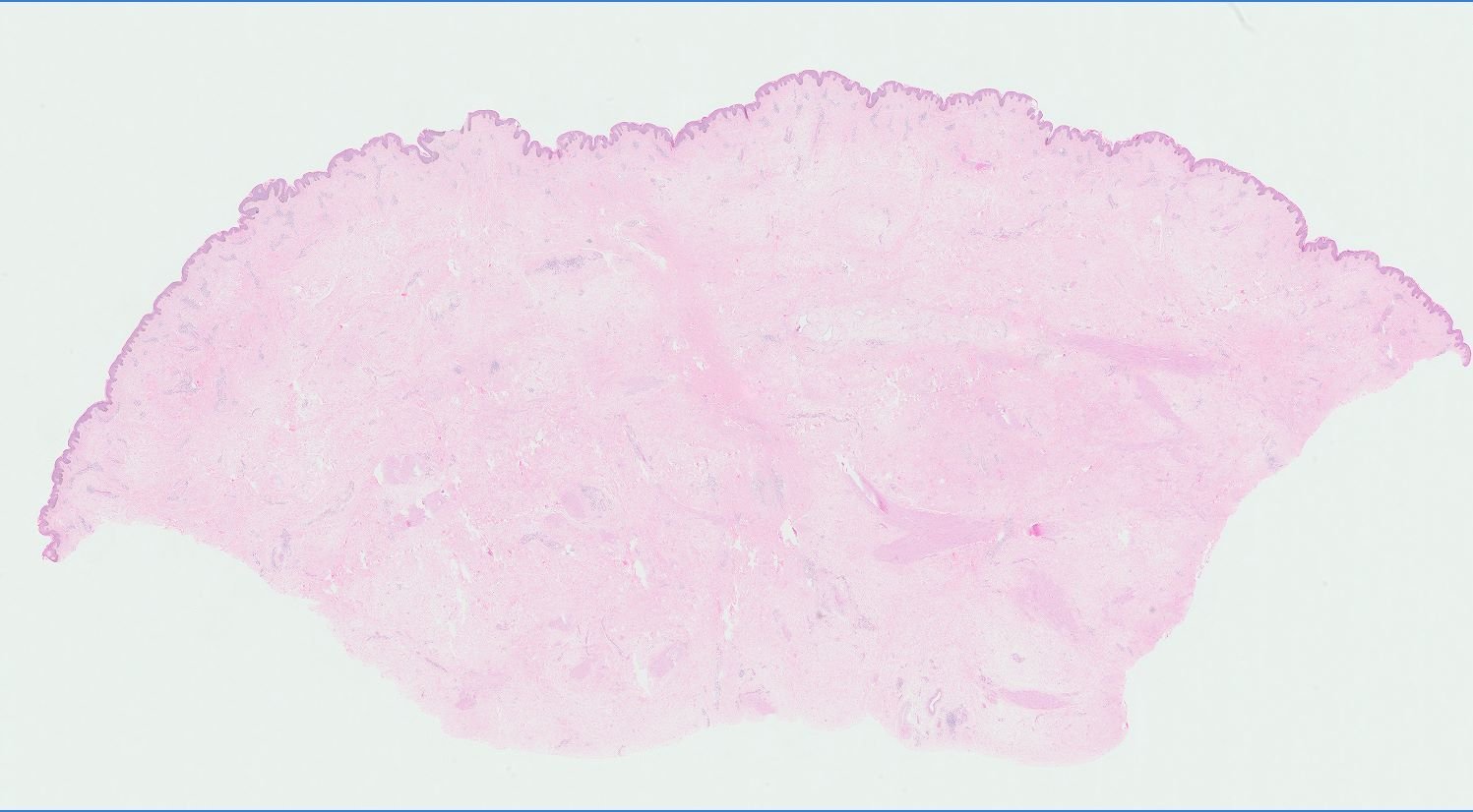
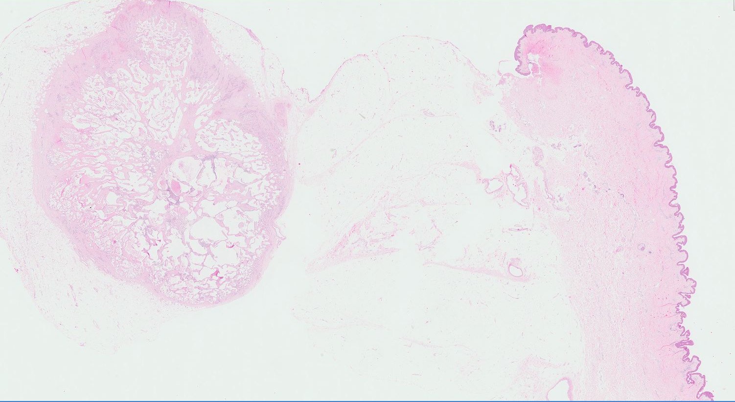
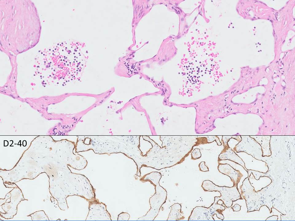
Microscopic findings: Extensive, marked stromal edema and areas of fibrosis were present. The grossly-identified nodule comprised a circumscribed proliferation of variably-sized vessels, containing luminal and stromal lymphocytes, lined by bland, mitotically-inactive endothelial cells highlighted by D2-40 immunohistochemical stain. No evidence of in-situ or invasive malignancy was present.
Diagnosis:
—